I swept the finished lightbox against both of its control inputs and measured its timing loop on a scope, then fitted the results.
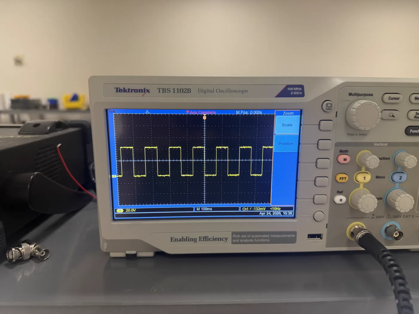
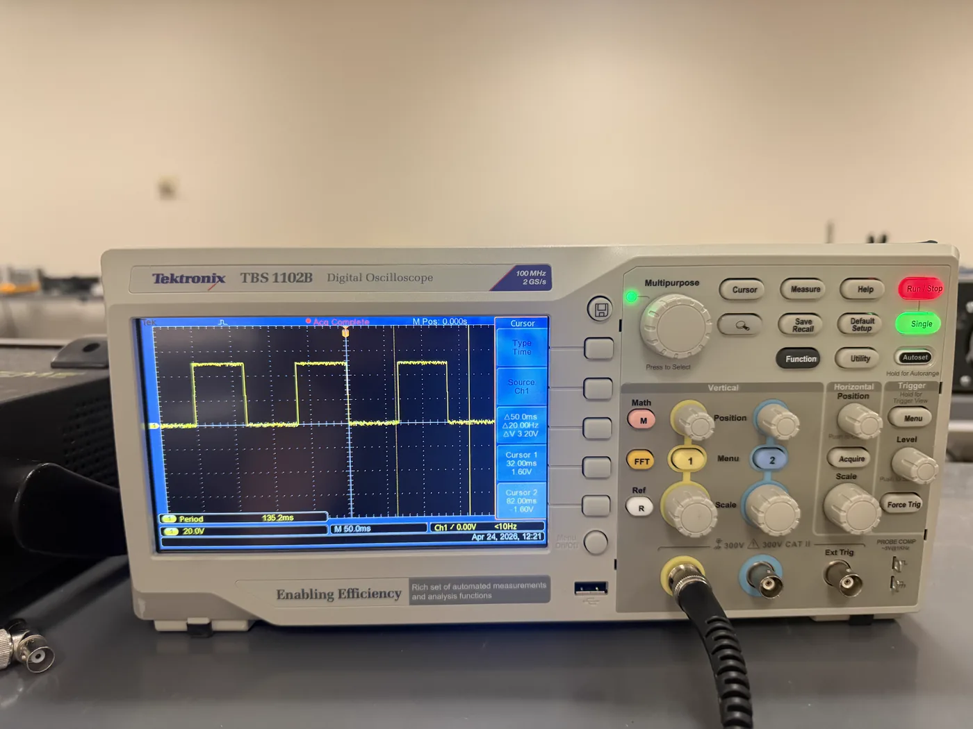
Switching output on the bench scope (Tektronix TBS 1102B), 100 ms/div.Time cursors placed on the output — measured period 135.2 ms.
Measured on the bench oscilloscope
Measurement
Sweep
Result
Eye brightness duty cycle vs control voltage
5 points · 0.862–3.3 V → 16.7–65.4 %
R² = 0.99994
Blink rate vs control voltage
5 points · 0–3.3 V → 5.00–7.35 Hz
R² = 0.974
Heartbeat period
10 trials
1000 ms, 10/10
ToolsOscilloscope · Curve fitting
Pacemaker — Strength–Duration Curve
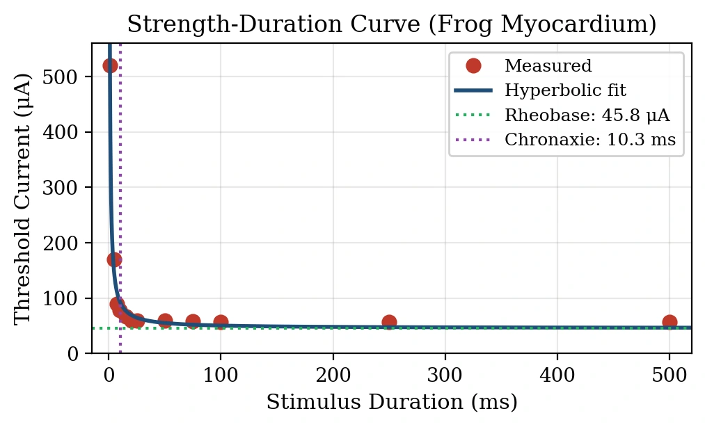
Threshold current measured against stimulus duration on frog ventricular myocardium, then fitted. The fit set the pulse width the pacemaker firmware uses.
Swept stimulus duration from 1 to 500 ms, recording the threshold current that evoked a visible ventricular contraction at each point.
Nonlinear least-squares fit to the hyperbolic form I(d) = b(1 + c/d): rheobase b = 45.8 µA, chronaxie c = 10.3 ms.
Firmware pulse width was then set to twice the measured chronaxie, taken from this fit.
Measured points with the hyperbolic fit — rheobase 45.8 µA, chronaxie 10.3 ms.
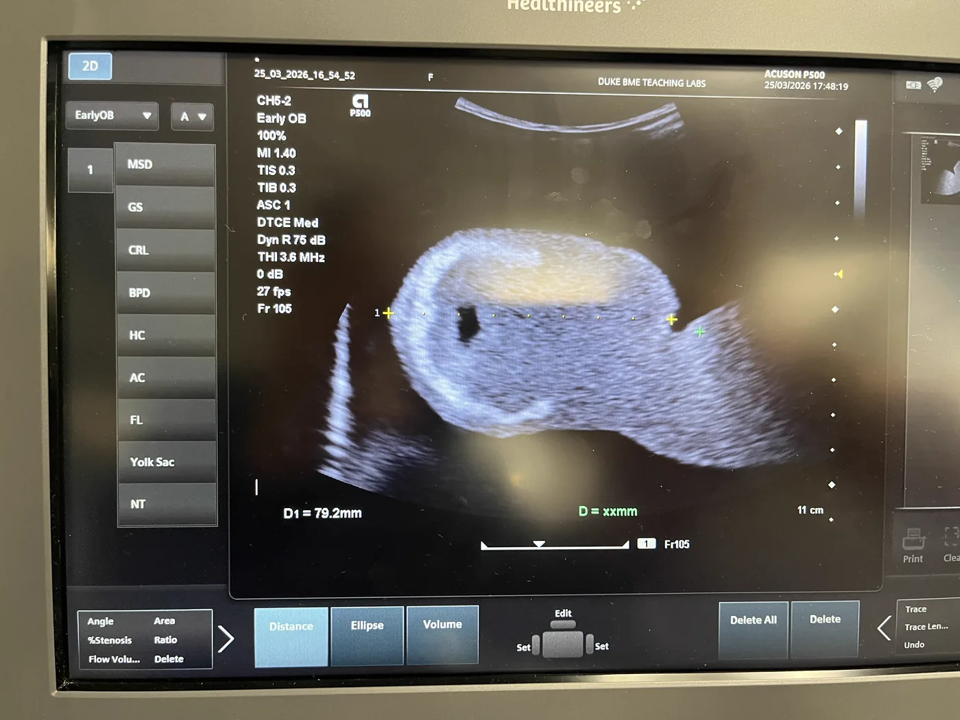
Scanning a training phantom on a clinical system: setting up the preset, optimizing the image, and placing measurement calipers.
Siemens Healthineers ACUSON P500, CH5-2 probe on the Early OB preset: MI 1.40, TIS/TIB 0.3, 75 dB dynamic range, tissue harmonics at 3.6 MHz, 11 cm depth.
Distance caliper placed across the target: D1 = 79.2 mm.
Acquired on a training phantom in a teaching lab, not on a patient.
ACUSON P500, Early OB preset — distance caliper reading D1 = 79.2 mm.